No-one with an interest in Mesozoic reptiles will have missed the week of controversy following Ibrahim et al.'s (2014) new reconstruction of Spinosaurus. The most important debate has focused on the allegedly reduced Spinosaurus hindlimbs, which are integral to the proposed locomotor and lifestyle hypotheses proposed for the 'new look' animal, but also difficult to reconcile with presented data. Scott Hartman, who's no stranger to producing high-quality skeletal reconstructions, blew this whistle first when he found the reconstructed proportions of the Spinosaurus neotype specimen - a series of vertebrae and hindlimb elements - were questionably scaled against measurements of the bones themselves. Lead author of the Spinosaurus study, Nizar Ibrahim, publicly responded and suggested that the measuring landmarks Scott used in comparing vertebral and hindlimb elements may be wrong. When reviewing the controversy before the weekend, I attempted my own scaling effort, using Nizar's suggested landmarks, but ended up replicating Scott's results almost exactly. I concluded "[s]omething - the original measurements of the specimen or the reconstruction - just doesn't add up, and I suspect the latter, as I figure someone would have owned up to and corrected simple numerical errors in the paper by now."
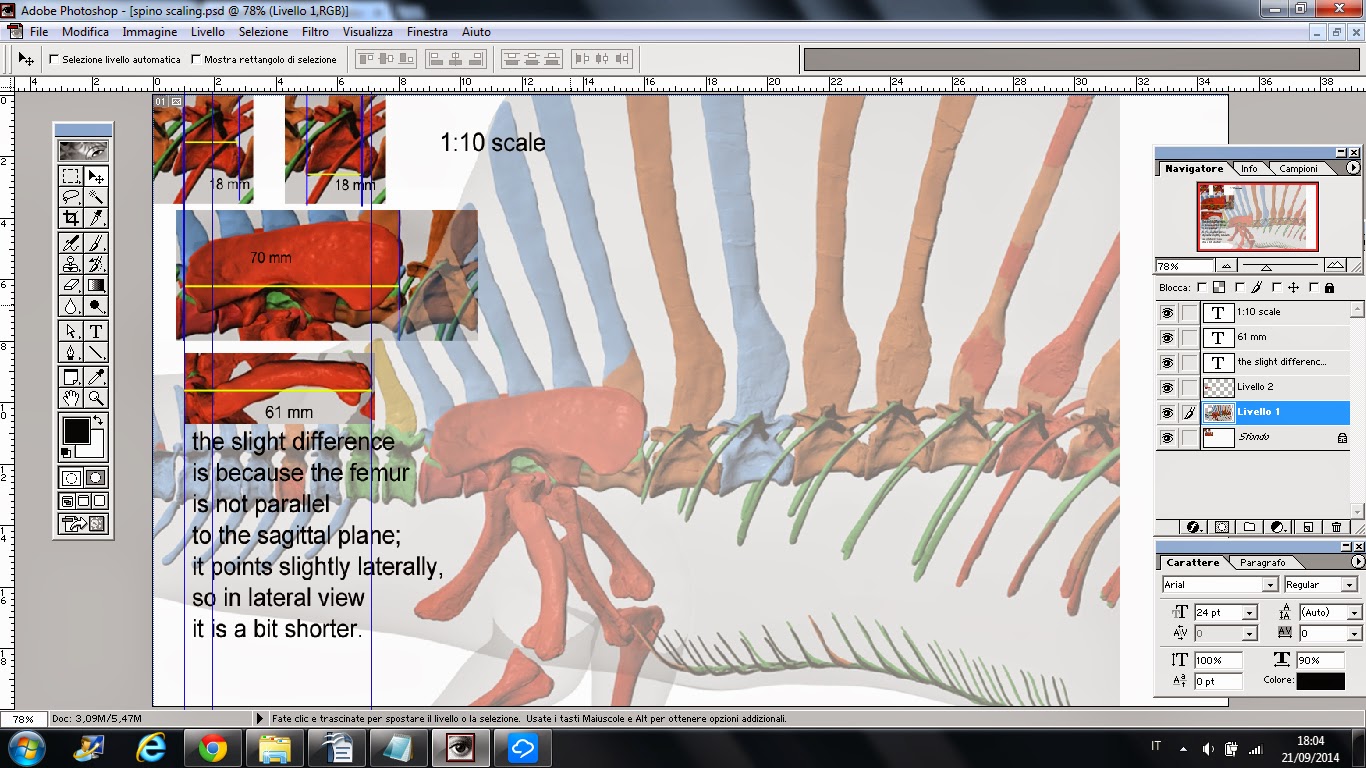
It turns out that I've got to eat a few of those words. Following my post, Nizar opened a chain of correspondence where I directly asked about these scaling issues. Nizar's response was bringing his coauthor Simone Maganuco into our chat, who had taken the time to demonstrate and describe how the restored vertebral and hindlimb lengths match the dimensions reported in the paper. In his screenshot and email, Simone provided an enlarged view of the restored Spinosaurus trunk and took the time to explain where he thought the alleged scaling errors came from. Appreciating their interest to a wide audience, Simone has kindly allowed me to reproduce his screengrab and email here.
Dear Mark,
It is nice to be in touch with you. I am writing to comment briefly on my photoshop image, forwarded by Nizar a couple of hours ago.
I hope it is the key to understand the misunderstanding about the measurements, so I would be really glad to know your opinion about it.
I have tried to replicate the coefficients for scaling obtained by you and Scott Hartman and here is my line of reasoning.
Look at the vertebra D8 in my photoshop image. For convenience, we can focus our attention on the D8 on the left.
The yellow line is 18 "units" (and matches our measurements in the table) but if you include the posteriormost margin of the slanted posterior face and the condyle you have nearly 23 units.
23:18=X:71 where 18 and 71 are also the measurements in cm in the table of the Science paper; 23 units is the length of the whole vertebra in the drawing; and X should be the length of the ilium to match the length of the vertebra in the drawing, if one assumes that the whole vertebra - and not the yellow line - is 18 units, i.e., if one thinks we used different landmarks and measured the maximum length of the centrum.
The value of X is 90.72 units.
90.72 /71 = 1.27 that is exactly the coefficient for pelvic girdle and hindlimb scaling suggested by Scott @ skeletaldrawing.com to resize the pelvis and the legs to match the size of the D8 vertebra measured with different landmarks (i.e., if 18 is considered the maximum length).
I can see that your coefficient is slightly lower, and I wonder if you have taken slightly lower measurements (it seems to be the case looking at the white lines in your test).
Do you think that this could be the explanation of what happened?
In the paper, we thought it was better to measure the vertebrae from rim to rim (the rounded margins of the faces), excluding the condyle, and at the same dorsoventral height (because some vertebrae are like parallelograms). It is easier to compare anterior dorsals and posterior dorsals in this way, and it is easier also to compare the centra with those of some specimens not prepared three-dimensionally but preserving well-articulated vertebrae, i.e. specimens in which it is difficult to look at the anterior condyle.
As what concerns the femur, it must be taken into account that there is also a slight perspective effect, because in the digital model it points a bit laterally. i.e., it is not 100% parallel to the sagittal plane.
The misunderstandings generated by the comparison between the figure and the table clearly indicate that we had to indicate our landmarks in one extra figure, or dedicate a couple of lines to this into the text to satisfy the need to compare figure and measurements by people who want to test our skeletal reconstruction.
When I work with palaeoartists to prepare illustrations and flesh-models I also compare figures and measurements, so I can understand this need.
Sometimes there are figures that are not 100% in the view indicated in the caption (also because it is not easy to put a bone in plane!) and sometimes it is difficult to understand the landmarks used to take measurements. What if I were in your shoes? Who knows... but I can understand that the new look of Spinosaurus has unexpected proportions that leads to think that there is something wrong.
In the monograph everything will be more clear because the detailed figures will report measurements directly on the bones, permitting everybody to see the landmarks.
In the meantime, however, I think it is useful to clarify this aspect.
Best wishes,
Simone
--
So there we have it: the measurements, landmarks and an image where they can be measured accurately. The latter is especially important because dorsal vertebra 8 in the full restoration is rather small, and thus prone to measuring errors even when measuring landmarks are known. A slip of a few pixels may not seem like much but, because the bone is a tiny component of a huge reconstruction, such minor errors can throw a scaling calibration right off. These risks were identified in Scott's originalposts, and it seems they have been borne out. Nevertheless, it is interesting that Scott and I - and others, according to some Facebook chat - found such similar results: this could be coincidence, or it might be that the published reconstruction lends itself to a erroneous interpretation. Either way, there is plenty of food for thought here as goes presentation and reading of reconstruction data. For the record, when attempting to replicate the scaling again, this time on the screenshot, I found my results matched measured values given in Ibrahim et al. (2014) within a few percent. My confidence in the published proportions is thus fully restored.
Hopefully this helps resolve the scaling controversy with the 'Spinosaurus reboot', and the result is much more confidence about the downright weird and remarkable anatomy of this genuinely unusual animal. Thanks to Nizar and Simone for taking the time to explain their work, and allowing me to post their response here.
It turns out that I've got to eat a few of those words. Following my post, Nizar opened a chain of correspondence where I directly asked about these scaling issues. Nizar's response was bringing his coauthor Simone Maganuco into our chat, who had taken the time to demonstrate and describe how the restored vertebral and hindlimb lengths match the dimensions reported in the paper. In his screenshot and email, Simone provided an enlarged view of the restored Spinosaurus trunk and took the time to explain where he thought the alleged scaling errors came from. Appreciating their interest to a wide audience, Simone has kindly allowed me to reproduce his screengrab and email here.
 |
| Image courtesy Nizar Ibrahim and Simone Maganuco, used with permission. |
It is nice to be in touch with you. I am writing to comment briefly on my photoshop image, forwarded by Nizar a couple of hours ago.
I hope it is the key to understand the misunderstanding about the measurements, so I would be really glad to know your opinion about it.
I have tried to replicate the coefficients for scaling obtained by you and Scott Hartman and here is my line of reasoning.
Look at the vertebra D8 in my photoshop image. For convenience, we can focus our attention on the D8 on the left.
The yellow line is 18 "units" (and matches our measurements in the table) but if you include the posteriormost margin of the slanted posterior face and the condyle you have nearly 23 units.
23:18=X:71 where 18 and 71 are also the measurements in cm in the table of the Science paper; 23 units is the length of the whole vertebra in the drawing; and X should be the length of the ilium to match the length of the vertebra in the drawing, if one assumes that the whole vertebra - and not the yellow line - is 18 units, i.e., if one thinks we used different landmarks and measured the maximum length of the centrum.
The value of X is 90.72 units.
90.72 /71 = 1.27 that is exactly the coefficient for pelvic girdle and hindlimb scaling suggested by Scott @ skeletaldrawing.com to resize the pelvis and the legs to match the size of the D8 vertebra measured with different landmarks (i.e., if 18 is considered the maximum length).
I can see that your coefficient is slightly lower, and I wonder if you have taken slightly lower measurements (it seems to be the case looking at the white lines in your test).
Do you think that this could be the explanation of what happened?
In the paper, we thought it was better to measure the vertebrae from rim to rim (the rounded margins of the faces), excluding the condyle, and at the same dorsoventral height (because some vertebrae are like parallelograms). It is easier to compare anterior dorsals and posterior dorsals in this way, and it is easier also to compare the centra with those of some specimens not prepared three-dimensionally but preserving well-articulated vertebrae, i.e. specimens in which it is difficult to look at the anterior condyle.
As what concerns the femur, it must be taken into account that there is also a slight perspective effect, because in the digital model it points a bit laterally. i.e., it is not 100% parallel to the sagittal plane.
The misunderstandings generated by the comparison between the figure and the table clearly indicate that we had to indicate our landmarks in one extra figure, or dedicate a couple of lines to this into the text to satisfy the need to compare figure and measurements by people who want to test our skeletal reconstruction.
When I work with palaeoartists to prepare illustrations and flesh-models I also compare figures and measurements, so I can understand this need.
Sometimes there are figures that are not 100% in the view indicated in the caption (also because it is not easy to put a bone in plane!) and sometimes it is difficult to understand the landmarks used to take measurements. What if I were in your shoes? Who knows... but I can understand that the new look of Spinosaurus has unexpected proportions that leads to think that there is something wrong.
In the monograph everything will be more clear because the detailed figures will report measurements directly on the bones, permitting everybody to see the landmarks.
In the meantime, however, I think it is useful to clarify this aspect.
Best wishes,
Simone
--
So there we have it: the measurements, landmarks and an image where they can be measured accurately. The latter is especially important because dorsal vertebra 8 in the full restoration is rather small, and thus prone to measuring errors even when measuring landmarks are known. A slip of a few pixels may not seem like much but, because the bone is a tiny component of a huge reconstruction, such minor errors can throw a scaling calibration right off. These risks were identified in Scott's originalposts, and it seems they have been borne out. Nevertheless, it is interesting that Scott and I - and others, according to some Facebook chat - found such similar results: this could be coincidence, or it might be that the published reconstruction lends itself to a erroneous interpretation. Either way, there is plenty of food for thought here as goes presentation and reading of reconstruction data. For the record, when attempting to replicate the scaling again, this time on the screenshot, I found my results matched measured values given in Ibrahim et al. (2014) within a few percent. My confidence in the published proportions is thus fully restored.
Hopefully this helps resolve the scaling controversy with the 'Spinosaurus reboot', and the result is much more confidence about the downright weird and remarkable anatomy of this genuinely unusual animal. Thanks to Nizar and Simone for taking the time to explain their work, and allowing me to post their response here.
Reference
- Ibrahim, N., Sereno, P. C., Dal Sasso, C., Maganuco, S., Fabbri, M., Martill, D. M., Zouhri, S. Myhrvold, N. & Iurino, D. A. (2014). Semiaquatic adaptations in a giant predatory dinosaur. Science, 1258750.