 |
| 1.5 Arsinotherium zitteli trotting about Eocene Egypt, looking a bit like they could be advertising farm products. But what's with those more elaborate than usual horns? |
Recently, I painted a portrait of Arsinoitherium for an upcoming book project and, based on my understanding of epidermal osteological correlates, I threw a keratinous sheath over the entire horn set (below). This is not a typical reconstruction - Arsinoitherium has been reconstructed with 'regular' mammalian skin (perhaps better termed 'villose skin' - Hieronymus et al. 2009) on its horns for decades but, as we all know, popularity and longevity don't always equal 'credibility' when it comes to fossil animal reconstructions.
 |
| Arsinoitherium zitteli, sporting antelope-like horn sheaths. |
It's difficult to turn away a good palaeobiological mystery, and because I like to make sure my work is as credible as it can be, I followed this question up with more research. I reasoned that the structure, development and surface texture of the three major types of mammalian headgear - horns, ossicones and antlers - could be compared to Arsinoitherium horns to see which, if any, is the best match and indicator of life appearance. Looking into this has been very informative and might be of interest to fellow palaeoartists as well as those interested in cool fossil animals, so I thought I'd share my thoughts and process here. We'll start by looking at Arsinoitherium horns themselves, then move through modern potential analogues, and finally compare them at the end to see which model seems most apt.
Arsinoitherium horns: growth, structure and surface texture
"...brought home by Dr. Andrews from Egypt, and after cleaning, strengthening, and the restoration of parts deficient on the left side by modelling from the right side, is now exhibited in the central hall of the Natural History Museum in Cromwell Road."Anonymous, 1903, p. 530
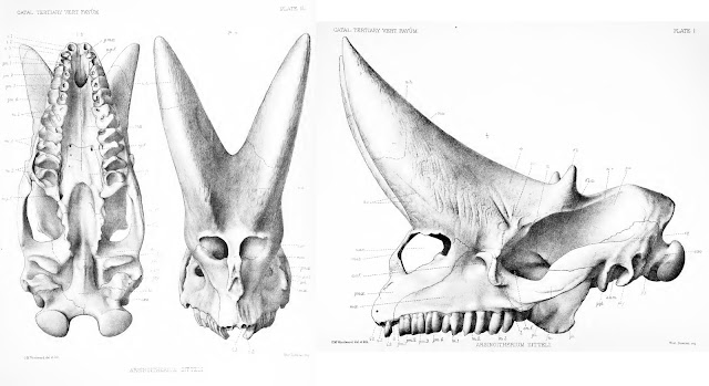
The fact that some parts of the skull were in less than stellar shape is evident from this photo of PV M 8463 (from the NHM's data portal): note the variation in colour and texture, reflecting places of reconstruction against real bone. Thus, while the familiar Arsinoitherium museum skull is a useful reference for morphology, illustrations and descriptions in technical literature will be more informative for reconstructing their integument. I've based my assessment mostly on Charles Andrews (1906) monograph, as well as that of Court (1992).
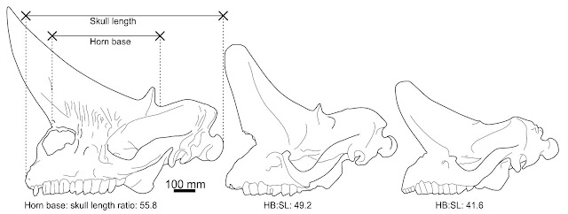
Structure. Both horn pairs of Arsinoitherium are relatively simple in gross shape and maintain the same basic morphology throughout their lives (below), though the horns of mature animals are wider, taller and more pointed than those of juveniles. The figures presented in Andrews (1906) show an increase in anterior horn base length from 41.6% in the smallest specimen to over 56% in the largest. Both horn sets are hollow, with vast internal cavities being supported by sheets of trabecular bone. In some places the exterior bone walls are surprisingly thin, only 5-10 mm (Andrews 1906).
Having learned something of the Arsinoitherium condition, let's take a look at how modern horns, antlers and ossicones compare...
Analogue 1. Bovid-style horns (keratinous sheaths over a bone core)
Bovid horns typify a widely used approach to cranial ornamentation and weaponry across Tetrapoda. They are perhaps the simplest approach to producing a sturdy cranial projection, being little more than a bony horn core covered in a hard keratinous sheath and are permanent feature in almost animals that bear them. The one exception is the pronghorn, which sheds its horn sheath annually (it also isn't a bovid). Biology, eh: can't we have one rule without an exception? |
| Bovid (bighorn sheep, Ovis canadensis) horn anatomy. From Drake et al. 2016. |
 |
| Schematic bovid horn growth, adapted from an illustration in Goss (2012). |
Analogue 2. Giraffe ossicones (skin over ossified dermis)
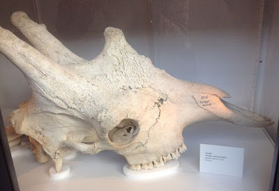
Giraffes have awesome skulls with two - and often more - ossicones that are covered in the same skin as the rest of their faces (Davis 2011). Their approach to cranial ornamentation seems unique to giraffes and their fossil relatives but might be an apt model for aberrant extinct forms, so is worth reviewing here. Clive Spinage (1968) provides an excellent overview of ossicone structure and development: the following is taken from his work.Structure. Ossicones are low humps or columnar protuberances, continuous with the surrounding skull anatomy but formed from dermal ossifications, not outgrowths of skull bones. They eventually fuse with the skull in adult life but, unlike the underlying skull bones, ossicones are solid and very dense - they are described as having 'ivory-like' in compactness and hardness by Spinage (1968). Mature specimens show increasingly complex shapes including development of swollen tips on the frontoparietal 'horns', as well as hornlets and bosses across the major 'forehead' ossicone. Having an adaptable, living integument is essential to this process, as the ossicone covering needs to change shape to reflect the changing size and complexity of the underlying bone.
 |
| Giraffe skulls are full of sinuses, but they do not extend to their ossicones, which are extremely dense. From Spinage (1968). |
 |
| Young adult male giraffe skull by Wikimedia user Nikkimaria, CC BY-SA 3.0. Note the flaky, irregular textures of the ossicones and their complex shape: they are much more intricate and developed than those of less mature animals. There's room for more irregularity and texture on this skull, too: the skulls of old males look like they have cathedral spires growing from their faces. |
Analogue 3. Deer antlers (bony projections atop cranial pedicles)
The familiarity of deer antlers allows us to forget what remarkable and unusual structures they are. Present almost universally in male deer (and in female reindeer), these elaborate, sometimes enormous structures are cast and regrown each year using a regenerative process that is the source of much anatomical and medical interest - no other mammal can regenerate such a complex appendage in this way, and the speed of the regeneration process is remarkable. Antlers are so unusual that they are only partly useful to our discussion here: we are primarily interested in antlers when they are covered in their velvet (specialised antler skin), as this is most comparable to the likely Arsinoitherium condition. Antler skin itself is interesting as, although it is continuous with the skin of the underlying pedicle, it lacks sweat glands and arrector pili (the tiny muscles that pull hair up or give us goosebumps) (Li and Suttie 2000). The antler pedicle (the permanent bony base) in contrast, is covered in the same type of skin as elsewhere on the body (Li and Suttie 2000). |
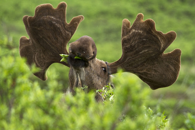
| A happy-looking moose (Alces alces) with his fuzzy antlers. Note the visible blood vessels on the underside of each palm. Photo by |
| More Alces antlers, this time without velvet. Note the long, branching channels. By Wikimedia user |
Surface texture. Antlers have variably developed rugosities consisting of conspicuous, long and branching channels impressed into smooth bone or around prominences and tubercles. These grooves are the impressions of blood and nervous networks that facilitated rapid antler growth. These textures are easily discerned even from a distance, and thus contrast with the texture of the pedicles, which are smoother and lined with relatively shallow, narrow and long impressions of vascular networks. It is unusual for hairy skin to leave such a significant osteological scar on underlying bone: typically, this form of epidermis leaves little to no remnant on skull bones (Hieronymus et al. 2009).
Arsinoitherium vs the analogues
Having looked at three major types of cranial projection in living animals, which - if any - best match the condition in Arsinoitherium?Giraffe ossicones are incomparable to Arsinoitherium horns in several aspects, perhaps the most significant being their increasing complexity and development of flaking bone textures in later life. Furthermore, the development of giraffe ossicones from bony growths in dermal tissues suggests a fundamentally different relationship between skull and dermis than of Arsinoitherium, where the bony horn component represents skull bones alone. There's enough differences here to question whether giraffe ossicones are a good model for the life appearance of Arsinoitherium horns.In being formed of polished, deeply vascularised bone, deer antlers are closer approximations of Arsinoitherium horns. However, there is so much weirdness associated with deer antler formation and tissues that they almost remove themselves from meaningful comparison to permanent skull horn cores. The fact that antler velvet, as hairy skin, is (to my knowledge) unique in leaving deep vascular channel impressions is a major issue here, implying that either antler bone is unusually susceptible to neurovascular imprinting (do they grow so fast that they grow around their blood vessels?) or that velvet is better at altering bone textures than other skin types. Both scenarios point to antlers having some endemic oddness about them, which complicates their use as a model for life appearance of non-antlered species.
All is not lost with the cervid data, however: antler pedicles are comparable to Arsinoitherium horns in being permanent outgrowths of bone, and they also have neurovascular impressions. However, these shallow grooves compare poorly to the deeper channels and pitting of Arsinoitherium horns. Indeed, there is little about antler pedicle texture to distinguish them from the surrounding skull bones, whereas the opposite is true for Arsinoitherium.
Our comparisons improve with the bovid horn condition, which seems to chime with the Arsinoitherium skull in many regards. Both are hollow outgrowths of skull bones supported by internal trabeculae; both have bone textures characterised by deep, bifurcating neurovascular channels as well as conspicuous longitudinal grooves and oblique foramina; and both maintain the same basic shape throughout growth - excepting some basic changes in base width and horn length. Further similarities include the development of particularly deep rugosties at the base of the horn cores, which is evident in at least large Arsinoitherium skulls (Andrews 1906). This interpretation is consistent with one of the longer (but still rather short, if we're honest) interpretations of the blood vessel impressions in Arsinoitherium:
"These channels evidently lodged blood-vessels which served for the conveyance of blood to or from the covering of the horn, and judging from the marked way in which both these vessels and those on the anterior face of the horns impress the bone, it seems probable that the covering was hard and of much, the same nature as that clothing the horn-cores of the cavicorn ruminants."C. Andrews (1906), p. 7
So...
Of the three models looked at here, it seems the basic structure and textural package of bovid-like horns best matches what we see in Arsinoitherium. Moreover, unlike the antler or ossicone models, there's no obvious mismatches with this configuration: pretty much everything we would correlate to a bovid-like horn anatomy seems present on or in the Arsinoitherium skull. The idea that a keratinous sheath might have existed in Arsinoitherium might seem odd, but it is not that outlandish given the apparent ease through which keratinous sheaths evolve. This is, after all, the tissue which has covered just about every claw, hoof, nail, horn, cranial dome and beak that has ever existed, whereas ossicones and antlers seem like specialised, clade-restricted approaches to cranial projections. The functionality of hollow Arsinoitherium horns is further reason to suspect a horn sheath. Studies of bovid horns suggest hollow cores and keratin sheaths compliment each other biomechanically, optimising the horns for for impact dissipation (Drake et al. 2016 and references therein). Stripped of a keratinous sheath, we find that hollow horn cores are great at transmitting energy but are brittle and prone to buckling and fracturing under heavy loading. It's only with a tough, fracture resistant keratin sheath that these structures can avoid breaking under heavy use so, if Arsinoitherium employed its horns for anything vaguely physically demanding, they probably needed a keratinous sheath.But - before we go crazy with this - do remember that the core of this analysis - the interpretation of Arsinoitherium headgear - is entirely literature based. I've not seen original specimens nor even modern, high-res imagery of an unreconstructed skull (this wasn't for lack of trying - the literature on these animals needs updating). Thus, while I've tried to be as thorough as I can with my observations, and as cautious as I can with my interpretations, I might be ignorant of some important detail. Take everything here with an appropriate pinch of salt, and please chime in below if you can provide superior insight. There's clearly scope for a more detailed study on this topic and, given how unique the horns of Arsinoitherium are, there might be some interesting functional findings to emerge from further investigation.
Enjoy monthly insights into palaeoart and fossil animal biology? Support this blog with a monthly micropayment, see bonus content, and get free stuff!
This blog is sponsored through Patreon, the site where you can help online content creators make a living. If you enjoy my content, please consider donating $1 a month to help fund my work. $1 might seem a meaningless amount, but if every reader pitched that amount I could work on these articles and their artwork full time. In return, you'll get access to my exclusive Patreon content: regular updates on research papers, books and paintings, including numerous advance previews of two palaeoart-heavy books (one of which is the first ever comprehensive guide to palaeoart processes). Plus, you get free stuff - prints, high quality images for printing, books, competitions - as my way of thanking you for your support. As always, huge thanks to everyone who already sponsors my work!References
- Andrews, C. W. (1906). A descriptive catalogue of the Tertiary Vertebrata of the Fayum. Publ. Brit. Mus. Nat. Hist. Land. XXXVII.
- Anonymous. (1903). A New Egyptian Mammal (Arsinoitherium) from the Fayûm. (1903). Geological Magazine, 10(12), 529-532.
- Court, N. (1992). The skull of Arsinoitherium (Mammalia, Embrithopoda) and the higher order interrelationships of ungulates. Palaeovertebrata, 22(1), 1-43.
- Davis, E. B., Brakora, K. A., & Lee, A. H. (2011). Evolution of ruminant headgear: a review. Proceedings of the Royal Society of London B: Biological Sciences, 278(1720), 2857-2865.
- Drake, A., Donahue, T. L. H., Stansloski, M., Fox, K., Wheatley, B. B., & Donahue, S. W. (2016). Horn and horn core trabecular bone of bighorn sheep rams absorbs impact energy and reduces brain cavity accelerations during high impact ramming of the skull. Acta biomaterialia, 44, 41-50.
- Goss, R. J. (2012). Deer antlers: regeneration, function and evolution. Academic Press.
- Hieronymus, T. L., Witmer, L. M., Tanke, D. H., & Currie, P. J. (2009). The facial integument of centrosaurine ceratopsids: morphological and histological correlates of novel skin structures. The Anatomical Record, 292(9), 1370-1396.
- Li, C., & Suttie, J. M. (2000). Histological studies of pedicle skin formation and its transformation to antler velvet in red deer (Cervus elaphus). The Anatomical Record, 260(1), 62-71.
- Osborn, H. F. (1907). Hunting the Ancestral Elephant in the Fayûm Desert: Discoveries of the Recent African Expeditions of the American Museum of Natural History. Century Company.
- Prothero, D. R., & Schoch, R. M. (2002). Horns, tusks, and flippers: the evolution of hoofed mammals. JHU press.
- Rose, K. D. (2006). The beginning of the age of mammals. JHU Press.
- Sanders, W. J., Kappelman, J., & Rasmussen, D. T. (2004). New large-bodied mammals from the late Oligocene site of Chilga, Ethiopia. Acta Palaeontologica Polonica, 49(3), 365-392.
- Spinage, C. A. (1968). Horns and other bony structures of the skull of the giraffe, and their functional significance. African Journal of Ecology, 6(1), 53-61.


